Chapter SummaryNeural Communication
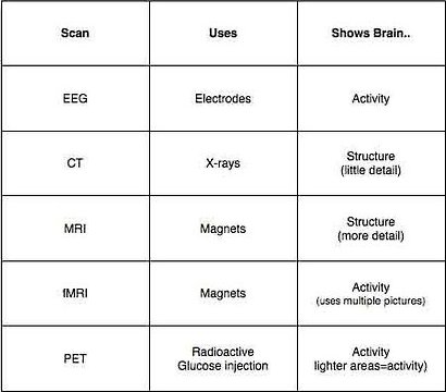
The body’s circuitry, the nervous system, consists of billions of individual cells called neurons. A neuron receives signals from other neurons through its branching dendrites and cell body, combines these signals in the cell body, and transmits an electrical impulse (the action potential) down its axon. When these signals reach the end of the axon, they stimulate the release of chemical messengers called neurotransmitters. These molecules pass on their excitatory or inhibitory messages as they traverse the synaptic gap between neurons and combine with receptor sites on neighboring neurons. Researchers are studying neurotransmitters to discern their role in behavior and emotion. Some drugs (agonists) excite by mimicking particular neurotransmitters or blocking their reuptake; others (antagonists) inhibit by blocking neurotransmitters. The Nervous System The central nervous system’s neurons in the brain and spinal cord communicate with the peripheral nervous system’s sensory and motor neurons. The peripheral nervous system has two main divisions. The somatic nervous system directs voluntary movements and reflexes. The autonomic nervous system, through its sympathetic and parasympathetic divisions, controls our involuntary muscles and glands. Like people clustering into neighborhoods, neurons cluster into working networks. The Endocrine System The endocrine system, one of the body’s communication systems, is a kindred system to the nervous system. Its glands release hormones at a slower rate than neurotransmitters, resulting in a longer lasting effect. The feeling outlasts the thought. However, the two systems are so closely interconnected that the distinction between them is sometimes difficult to decipher. The Brain The Tools of Discovery Clinical observations have long revealed the general effects of damage to various areas of the brain. But CT and MRI scans now reveal brain structures, and EEG, PET, and functional MRI recordings reveal brain activity. By surgically lesioning or electrically stimulating specific brain areas, by recording the brain’s surface electrical activity, and by displaying neural activity with computer-aided brain scans, neuroscientists explore the connections among brain, mind, and behavior. Older Brain Structures The brainstem begins where the spinal cord swells to form the medulla, which controls heartbeat and breathing. Within the brainstem, the reticular formation controls arousal. Atop the brainstem is the thalamus, the brain’s sensory switchboard. The cerebellum, attached to the rear of the brainstem, coordinates muscle movement. Between the brainstem and cerebral cortex is the limbic system, which is linked to memory, emotions, and drives. One of its neural centers, the amygdala, is involved in responses of aggression and fear. Another, the hypothalamus, is involved in various bodily maintenance functions, pleasurable rewards, and the control of the hormonal system. The Cerebral Cortex Each hemisphere of the cerebral cortex—the neural fabric that covers the hemispheres—has four geographic areas: the frontal, parietal, occipital, and temporal lobes. Small, well-defined regions within these lobes control muscle movement and receive information from the body senses. However, most of the cortex—its association areas—is uncommitted to such functions and is therefore free to process other information. Some brain regions serve specific functions. The brain divides its labor into specialized subtasks and then integrates the various outputs from its neural networks. Thus, our emotions, thoughts, and behaviors result from the intricate coordination of many brain areas. Language, for example, depends on a chain of events in several brain regions. If one hemisphere is damaged early in life, the other will pick up many of its functions, thus demonstrating the brain’s plasticity. The brain becomes less plastic later in life. Frequently, however, nearby neurons can partially compensate for damaged ones, as when a person recovers from a stroke or brain injury. Our Divided Brain Clinical observations long ago revealed that the left cerebral hemisphere is crucial for language. Experiments on people with a severed corpus callosum have refined our knowledge of each hemisphere’s special functions. Separately testing the two hemispheres, researchers have confirmed that in most people the left hemisphere is the more verbal, and that the right hemisphere excels in visual perception and the recognition of emotion. Studies of healthy people with intact brains confirm that each hemisphere makes unique contributions to the integrated functioning of the brain. Key TermsBiological Psychology - a branch of psychology concerned with the links between biology and behavior.
Neuron - a nerve cell; the basic building block of the nervous system. Dendrite - the bushy, branching extensions of a neuron that receive messages and conduct impulses toward the cell body. Axon - the extension of a neuron, ending in branching terminal fibers, through which messages pass to other neurons or to muscles or glands. Myelin - a layer of fatty tissue segmentally encasing the fibers of many neurons; enables vastly greater transmission speed of neural impulses as the impulse hops from one node to the next. Action Potential - a neural impulse; a brief electrical charge that travels down an axon. the action potential is generated by the movement of positively charged atoms in and out of channels in the axon's membrane. Threshold - the level of stimulation required to trigger a neural impulse. Synapse - the junction between the axon tip of the sending neuron and the dendrite or cell body of the receiving neuron. The tiny gap at this junction is called the synaptic gap or cleft. Neurotransmitters - chemical messengers that traverse the synaptic gaps between neurons. When released by the sending neuron, neurotransmitters travel across the synapse and bind to receptor sites on the receiving neuron, thereby influencing whether that neuron will generate a neural impulse. Acetylcholine (ACh) - a neurotransmitter that enables learning and memory and also triggers muscle contraction. Endorphins - "morphine within"--natural, opiate-like neurotransmitters linked to pain control and to pleasure. Nervous System - the body's speedy, electro-chemical communication system, consisting of all the nerve cells of the peripheral and central nervous systems. Central Nervous System (CNS) - the body system composed of the brain and the spinal cord. Peripheral Nervous System (PNS) - the sensory and motor neurons that connect the central nervous system to the rest of the body. Nerves - neural "cables" containing many axons. These bundled axons, which are part of the peripheral nervous system, connect the central nervous system with muscles, glands, and sense organs. Sensory Neurons - neurons that carry incoming information from the sense receptors to the central nervous system. Motor Neurons - neurons that carry outgoing information from the central nervous system to the muscles and glands. Interneurons - central nervous system neurons that internally communicate and intervene between the sensory inputs and motor outputs. Somatic Nervous System - the division of the peripheral nervous system that controls the body's skeletal muscles. Also called the skeletal nervous system. Autonomic Nervous System - the part of the peripheral nervous system that controls the glands and the muscles of the internal organs (such as the heart). Its sympathetic division arouses; its parasympathetic division calms. Sympathetic Nervous System - the division of the autonomic nervous system that arouses the body, mobilizing its energy in stressful situations. Parasympathetic Nervous System - the division of the autonomic nervous system that calms the body, conserving its energy. Reflex - a simple, automatic, inborn response to a sensory stimulus, such as the knee-jerk response. Neural Networks - interconnected neural cells. With experience, networks can learn, as feedback strengthens or inhibits connections that produce certain results. Computer simulations of neural networks show analogous learning. Endocrine System - the body's "slow" chemical communication system; a set of glands that secrete hormones into the bloodstream. Hormones - chemical messengers, mostly those manufactured by the endocrine glands, that are produced in one tissue and affect another. Adrenal Glands - a pair of endocrine glands just above the kidneys. the adrenals secrete the hormones epinephrine (adrenaline) and norepinephrine (noradrenaline), which help to arouse the body in times of stress. Pituitary Glands - the endocrine system's most influential gland. Under the influence of the hypothalamus, the pituitary regulates growth and controls other endocrine glands. Lesion - tissue destruction. A brain lesion is a naturally or experimentally caused destruction of brain tissue. Electroencephalogram (EEG) - an amplified recording of the waves of electrical activity that sweep across the brains surface. Waves are measured by electrodes placed on the scalp Positron Emission Tomography (PET) - a visual display of brain activity that detects where a radioactive form of glucose goes while the brain performs a given task. Magnetic Resonance Imaging (MRI) - a technique that uses magnetic fields and radio waves to produce computer generated images that distinguish among different types of soft tissue; allows us to see structures within the brain. Functional Magnetic Resonance Imaging (fMRI) - a technique for revealing blood flow and, therefore, brain activity by comparing successive MRI scans. MRI scans show brain anatomy; fMRI scans show brain function. Brainstem - the oldest part and central core of the brain, beginning where the spinal cord swells as it enters the skull; the brainstem is responsible for automatic survival functions. Medulla - the base of the brainstem; controls heartbeat and breathing. Reticular Formation - a nerve network in the brainstem that plays an important role in controlling arousal. Thalamus - the brain's sensory switchboard, located on top of the brainstem; it directs messages to the sensory receiving areas in the cortex and transmits replies to the cerebellum and medulla. Cerebellum - the "little brain" attached to the rear of the brainstem; its functions include processing sensory input and coordinating movement output and balance. Limbic System - a doughnut-shaped system of neural structures at the border of the brainstem and cerebral hemispheres; associated with emotions such as fear and aggression and drives such as those for food and sex. Includes the hippocampus, amygdala, and hypothalamus. Amygdala - two lima bean-sized neural clusters that are components of the limbic system and are linked to emotion. Hypothalamus - a neural structure lying below the thalamus; directs eating, drinking, body temperature; helps govern the endocrine system via the pituitary gland, and is linked to emotion. Cerebral Cortex - the intricate fabric of interconnected neural cells that covers the cerebral hemispheres; the body's ultimate control and information-processing center. Glial Cells - cells in the nervous system that support, nourish, and protect neurons. Frontal Lobes - the portion of the cerebral cortex lying just behind the forehead; involved in speaking and muscle movements and in making plans and judgments. Parietal Lobes - the portion of the cerebral cortex lying at the top of the head and toward the rear; receives sensory input for touch and body position. Occipital Lobes - the portion of the cerebral cortex lying at the back of the head; includes the visual areas, which receive visual information from the opposite visual field. Temporal Lobes - the portion of the cerebral cortex lying roughly above the ears; includes the auditory areas, each of which receives auditory information primarily from the opposite ear. Motor Cortex - an area at the rear of the frontal lobes that controls voluntary movements. Sensory Cortex - the area at the front of the parietal lobes that registers and processes body touch and movement sensations. Association Areas - areas of the cerebral cortex that are not involved in primary motor or sensory functions; rather, they are involved in higher mental functions such as learning, remembering, thinking, and speaking. Aphasia - impairment of language, usually caused by left hemisphere damage either to Broca's area (impairing speaking) or to Wernicke's area (impairing understanding). Broca's Area - controls language expression-an area of the frontal, usually in the left hemisphere, that directs the muscle movements involved in speech. Wernicke's Area - controls language reception-a brain area involved in language comprehension and expression;usually in the left temporal lobe. Plasticity - the brain's capacity for modification, as evident in brain reorganization following damage (especially in children) and in experiments on the effects of experience on brain development. Corpus Callosum - the large band of neural fibers connecting the two brain hemispheres and carrying messages between them. Split Brain - a condition in which the two hemispheres of the brain are isolated by cutting the connecting fibers (mainly those of the corpus callosum) between them. |
People To Know*Phineas Gage - railroad worker who had a piece of pipe blow a hole through his head; survived by his personality changed; scientists learned you could manipulate the brain.
Candace Pert and Solomon Snyder - discovered the body produces its own morphine by injecting laboratory animals with morphine. *Joseph Gall - founder of phrenology (thought that characteristics could be determined by bumps on the skull). Oliver Sacks - neurologist who writes books containing interesting stories about his patients (An Anthropologist from Mars; The Man who Mistook his Wife for a Hat). Sandra Witelson - gained access to the brain of Albert Einstein and discovered that a region used for mathematical thinking was 15% wider than the average brain; theorized that either Einstein was born with a gifted brain or that it grew because he used that area of his brain more often. Hans Burger - invented a machine that could detect, amplify, and record waves of electrical activity in the brain using metal electrodes pasted to the surface of the scalp (EEG). Heinrich Kluver and Paul Bucy - proved that lesions in the temporal lobe (including the amygdala) calmed ferocious monkeys. Vernon Mark - Neurosurgeon who implanted electrodes in an overly aggressive patient's (Julia) brain and recorded the activity of the amygdala to discover if it was making her violent; discovered that high activity in the amygdala was causing violence; Mark performed surgery and destroyed part of her amygdala and caused her fits of rage to go away. Julia - Overly aggressive patient who suffered from severe fits of rage; Vernon Mark performed surgery to destroy some of her amygdala and lost most of her violent tendencies. Wilder Penfield - stimulated exposed parts of the cortex during surgery on his epileptic patients and mapped the human cortex in 1947; discovered that certain areas of the brain specialize in receiving sensory information. *Paul Broca - French physician who observed people who have incurred damage in an area of the frontal lobe of the left hemisphere lose the ability to form words to produce fluent speech; region of the brain called Broca's area. *Carl Wernicke - German neurologist who found that people with damage to a part of the left temporal lobe lose their ability to comprehend speech; region of the brain called Wernicke's Area. Gustav Fechner - German physicist who proposed that each side of the brain has its own mind; speculated if the brain was divided in half, you would have two separate streams of consciousness. Philip Vogel and Joseph Bogen - Two neurosurgeons who described the case of a severe epileptic man who had a split-brain procedure done; his behavior seemed normal at first, but after further research it was realized that there were side effects. Roger Sperry - conducted a study in 1968 showing that the two halves of the brain work together; took split-brain patients (client named NG) and had them focus on a black dot while occasionally flashing a picture of a spoon; when spoon was shown in the right hemisphere NG could say spoon because it went to the left brain; when spoon was shown in the left hemisphere NG couldn't say spoon but could pick it out because it went to the right brain; Sperry said in split brain subject the two sides function independently, but in normal brains the two sides work together. Micheal Gazzaniga - worked with Sperry; conducted a similar experiment to Sperry using the word tea cup instead. Jerre Lecy - took pictures of various faces and cut them and pasted different halves together; had patients stare at different halves of the picture and report who they saw. Justine Sergent - explored the issue of information traveling through split brain patients through "subcortical" structures. Sir John Eccles - disagreed with Sperry's theories of split brain patients having two different minds. Joseph Ledoux - tested a split brain patient known as PS who had teh ability to communicate with the right hemisphere by arranging scrabble letter with the left hand, however the two sides of his brain often disagreed with each other. Mark Rosenzweig - built an amusement park for rats to examine the effects of an enriched environment on neural development; proved experiences can spark the growth of new synaptic connections and mold the brain's neural architecture. Avi Karni and Leslie Ungerleider - tested the proposition that repeated stimulation of a body part would cause corresponding changes in the human brain; showed that repetition can spark the buildup of a new synaptic connection among neurons. Michael Merzenich - severed the nerve of the middle finger of an adult monkey and found that the area of somatosensory cortex dedicated to that finger did not wither away, but that nearby neurons activated by other fingers filled in the dormant region. V.S. Ramachandran - author of Phantoms in the Brain; wrote about phantom pains in amputees based off his experience with teenage soccer player, Tom Sorenson, who lost his left arm in an auto accident; stroked Tom's face with a Q-tip and discovered different areas of the face stimulated different areas on his missing arm; "mapped" the missing arm in the boys face; discovered some nerves are interconnected. * Most important of the people PSYCH SIM 5 ACTIVITIESTricky Spots
Extras
Videos | ||||
